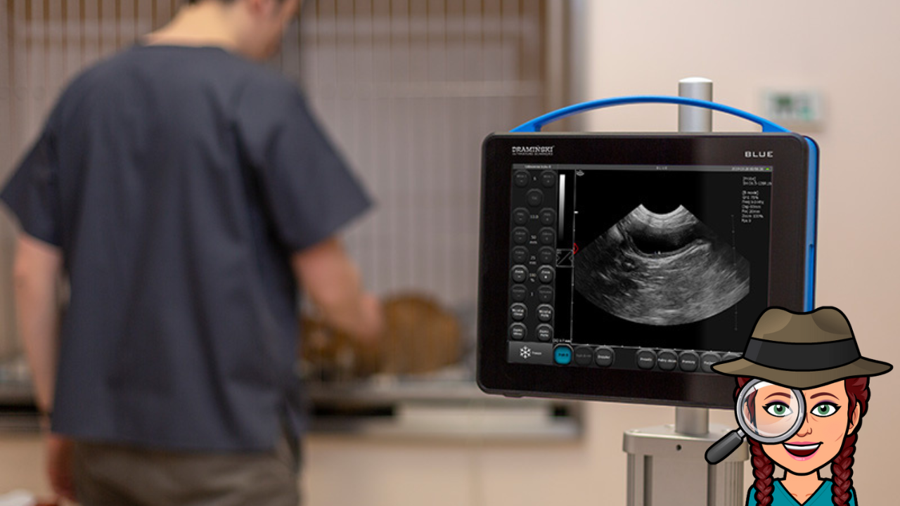
3 Mistakes Veterinary Professionals Make With Grey Scale Images
Dec 19, 2022
Have you ever asked the questions…
- What are grey scale images in ultrasound?
- How do I understand grey scale images?
If you have, then this blog post will highlight the 3 mistakes people often make with grey scale images so you can progress and confidently understand your patient's grey scale images.
The term Echogenicity is used when describing ultrasound images, and it refers to the number of sound waves that bounce back to the probe where tissue interfaces change. This includes sound waves that aren't attenuated, reflected, refracted, or scattered.
The stronger the reflection, the brighter the dot we see on the image. And grey scale refers to the scale from black to white that an image can produce. However, what exactly are the three most frequent mistakes that veterinary professionals make when working with grey scale images?
1) Understanding echogenicity is relative to surrounding tissue echogenicity.
When we discuss the echogenicity of an organ, our interpretation of that value will be wholly determined by the tissue surrounding the organ. When compared to the cortex of the kidney, the liver has a hyperechoic echogenicity, but when compared to the spleen, the liver has a hypoechoic echogenicity. This is one example of the relationship between the echogenicity of different organs.
A useful mnemonic is My Cat Loves Sunny Places to remember the order of organs echogenicity from hypoechoic (darker grey) to hyperechoic (brighter/whiter grey) – renal Medulla and Cortex, Liver, Spleen, Prostate.
2) Artefacts can influence echogenicity.
Mineralised areas such as bone and calculi reflect most of the ultrasound waves at their surface interface. Therefore this appears as white on the screen with black (acoustic shadow) beyond where no ultrasound waves penetrate.
Ultrasound waves pass easily through fluid without attenuating (losing energy) or returning waves to the probe – this means that it appears black (anechoic) on the screen.
But because the sound waves have not attenuated when passing through fluid, those sound waves reflecting from soft tissue beyond the fluid appear hyperechoic relative to sound waves which have passed the same distance but through soft tissue (acoustic enhancement).
3) Overall Gain can Effect Our Interpretation of Echogenicity.
When we adjust our overall gain, the image on our screen appears brighter or darker. What is actually happening is that we are amplifying the returning sound waves. If we adjust the overall gain during an examination of an abdomen multiple times, we will struggle to compare a liver’s echogenicity (when the overall gain might be set higher) to a spleen (when perhaps the overall gain was set lower).
Try to wait until the gel has absorbed (before it is fully absorbed, the image is generally dark, and it is tempting to dial up the overall gain), then set the overall gain for a large organ such as the liver and then don’t adjust for the rest of the abdomen.
To further support you, here are some other key terms to understand grey scale images:
- Hyperechoic refers to a tissue which produces a strong reflection of sound waves back to the transducer, and this appears white.
- Hypoechoic refers to weak returning sound waves, and these will appear grey on the B-mode ultrasound screen.
- Anechoic refers to an area where no sound waves are returning to the probe – this may be a property of the tissue itself or because a hyperechoic tissue has caused reflection of all the sound waves before they had a chance to reach this area.
- Homogenous is when there is a uniform shade of grey throughout an organ, whereas Heterogenous describes non-uniform shades of grey.
Having the confidence and know-how to perform veterinary ultrasound is such a valuable tool. In addition to being able to differentiate between fluid and solid tissue, it offers a wealth of information regarding the internal structure of the organs.
So, to get the most out of your scanning techniques, try to practise as much as you can so that you have a solid understanding of grey scale images.
In my FOVU Club membership, I cover topics such as grey scale imagery, case studies, and monthly focus sessions on different topics and organs, so if you feel as though you need more information and support, you can take advantage of those resources. Doors open to new members in January, so add your name to the FOVU Club interested list by clicking here to be notified of this date and learn more.
You can also join my 'Get Started With Veterinary Ultrasound 5-day Challenge,' which begins on Monday, January 16th, 2023. By clicking here, you can get the accountability you need to start using your ultrasound machine and developing your ultrasound skills and knowledge.
